- Aural atresia skin#
- Aural atresia free#
“These children are born with a moderate to severe hearing loss, and after surgery, many times I can bring their hearing into the normal range,” says neurotologist Bradley Kesser, MD, a professor in UVA’s Department of Otolaryngology - Head and Neck Surgery. For children born with aural atresia – the lack of a fully developed ear canal and middle ear in one or both ears – UVA’s aural atresia repair program offers hope for a normal life.
Case no 2 is An 18 year-old female patient referred to our institute with painful post-auricular discharging sinus on her left side associated with swelling and redness around it.ENT Innovations, Innovations & Excellence 2021-22, Pediatric Excellence įrom birth, the ability to hear is essential for language and speech development. the boy is scheduled to have his 1st audiogram in 3rd week postoperatively. here the patient is seen in the 2nd week showing granulating wound with healthy pedicle skin.silver nitrate was applied to the granulations and EAC was packed with antibiotic impregnated gelfoam. The patient was discharged on the 2nd postop day and sutures were removed on 1 postop week. Following proper placement of the meatus the external ear was stabilized with subcutaneous sutures and ear canal was packed with spongiston wicks and BIPP packs.mastoid dressing was applied. a “U”-shaped pedicle flap hinged at the tragal remanent was then positioned into the new ear canal and sutured to a cuff of periosteum. fascia graft was placed directly on the ossicular Mass so as it was Approximately in center of the new tympanic membrane. peripheral bone was then drilled to gain room for the placement of the fascial graft. Normal mobile stapes suprastructure was confirmed by gentle palpation. facial nerve was seen having a short vertical segment while making a sharp curve anteriorly. diamond bur was Used to thin the atretic plate to eggshell thickness and gently picked away in small pieces. fused malleus head and incus body were found to be mobile on palpation. body of the incus was identified and confirmed by gentle Palpation. fused incus-malleus complex was encountered At a depth of approx 1.5 cm. Dense atretic bone was found and followed medially. Drilling was done by staying superior and anteriorly.Care was taken to hug the tegmen and the glenoid fossa.  The temporal root of the zygomatic arch and the glenoid fossa were identified and the cribriform area of the mastoid process was used as a landmark for drilling. An endaural incision is made and Soft tissue was elevated off the mastoid process in a posterior to anterior direction. A 1/2-inch swath of hair is shaved around the external ear
 Aural atresia skin#
lateral face and the skin graft donor site were prepared and draped.

Associated risks of total deafness and facial nerve paralysis were explained to the parents.
The boy was graded 9 on Jahrsdoerfer system and planned for surgery of the right ear. Ct san revealed partially canalised right external auditory canal with aerated middle ear and intact ossicles where as left middle ear showed abnormal configuration with no definitive ossicles. speech therapist suggest adequate receptive and expressive language with good attention span. Aural atresia free#
free field hearing test reveal 60 and 80 db in right and left ear respectively.the boy had been wearing soft band bone conducting hearing aid since birth and studying in a special education school.
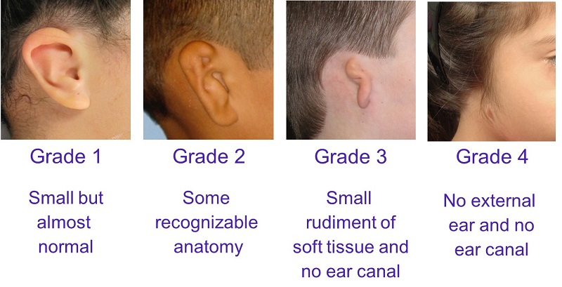
BERA revealed the boy having 60 and 70 db hearing threshold in right and left ear respectively. on examination altman grade 3 aural atresia with marx grade 3 microtia were seen bilaterally.  Case no 1 is A 6 year old boy brought to our institute by his consanguineous parents for scheduled ear surgery for bilateral hearing loss since birth. I dr zeeshan resident in ENT head and neck surgery department CMH rwp will be presenting two cases of congenital aural atresia with different surgical scenarios. Respected teachers and my fellow colleagues AOA. Oliver ER, Hughley BB, Shonka DC, Kesser BW.Revision aural atresia surgery: indications and outcomes. Nishizaki K, Masuda Y, Karita K.Surgical management and its post-operative complications in congenital aural atresia.Acta Congenital absence of the oval window: diagnosis, surgery, and audiometric
De Alarcon A, Jahrsdoerfer RA, Kesser BW.


 0 kommentar(er)
